1H/X and 1H/19F MRI Volume Coils
Mouse Coils – Rat Coils – Rabbit Coils -Primate Coils – Custom Coils
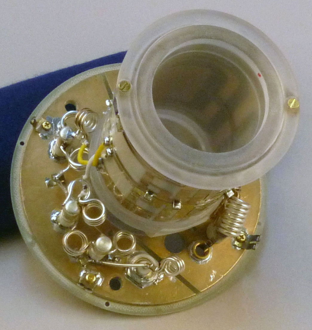
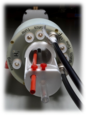
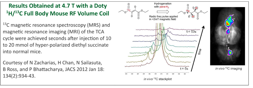
Dual-frequency MR Coils enable MR spectroscopy and imaging techniques, including hyperpolarized nuclei. Detailed full-wave simulations ensure the highest sensitivity, homogeneity, and isolation between channels. B1 field maps and SAR maps included with each coil. Flexible design can be customized for your application and MR system.
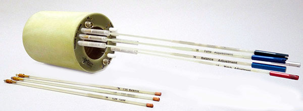
- Efficient, easy to tune and match over a broad range of sample loadings.
- Each channel for TxRx.
- For observe / decouple – with both channels simultaneously.
- For interleaved acquisitions – with each channel used sequentially.
- Robust design, mechanical stability.
A Dual Frequency Mouse Brain Volume coil (right) for 15.2 T imager with 6 cm bore. The high frequency (650 MHz 1H) and tight spacing made this coil quite challenging. A 36 mm ID x 35 mm FOV allows a surface coil receive, when desired. Tuning included: 1H-19F and multi-X with a low gamma nucleus: 23Na,13C, 2H, 17O, 15N, and 14N.
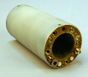
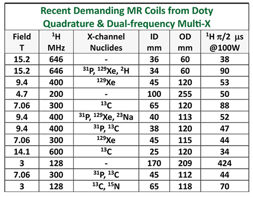
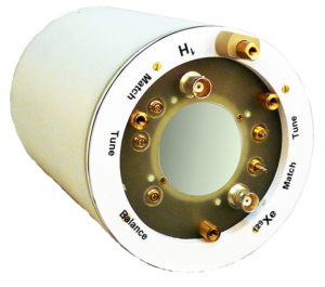
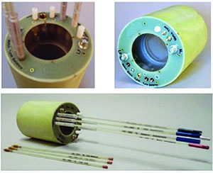 65 x52 mm 1H/X Dual Freq Coils | Examples of dual-frequency coils we have supplied (ID x RF length): 65 x 52 mm, 1H/X, (X=13C, 15N),@ 3 T; 1.5 T; 1 T; 0.5 T; and 0.3 T. 45 x 36 mm, 1H/X (X=31P, 13C), @ 7 T. 38 x 34 mm, 1H/X, (X=31P, 13C), @ 9.4 T. 25 x 22 mm, 1H/X, (X=31P, 13C), @ 9.4 T. 25 x 22 mm, 1H/13C, @ 1 T. 200 x 160 mm, 1H/23Na, @7 T. 38 x 55 mm, 1H/19F, @ 7 T. 32 x 25mm, 1H/19F, @9.4T |
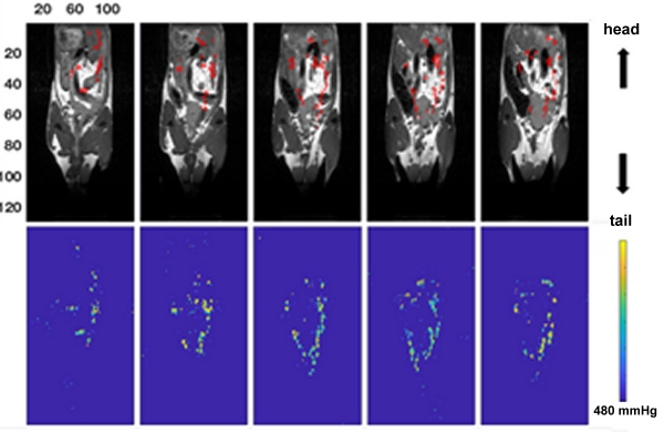
Mouse Images Obtained with a Doty 1H/19F 38 mm Volume Coil
Pantom Images from a Dual Frequency 1H/19F 32 mm Volume Coil.

