> Rat Spinal Cord Imaging on Unique Imaging Coils
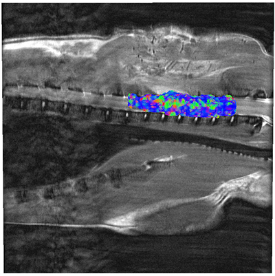
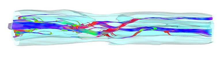
A customer contacted us with a request to build custom coils for their studies on rat spinal cord, both in vivo and ex vivo, motivated by performance issues related to RF homogeneity and shielding exhibited in current OEM coils. A quadrature volume coil with 56 mm ID was constructed for the in vivo imaging (on awake or mildly anesthetized rats), and was utilized at the study onset. The volume coil’s excellent sensitivity made it possible to generate high resolution DTI images of an injured spinal cord in vivo, with weekly scanning to monitor treatment progress. When treatments ended, it was desired to image an excised cord, with smaller fov to visualize more specific spinal tracts. High S/N and excellent B1 homogeneity were top priorities. To meet this goal, a one-turn RF saddle coil was used to construct a small coil for the spinal cord contained in a 5 mm NMR tube.
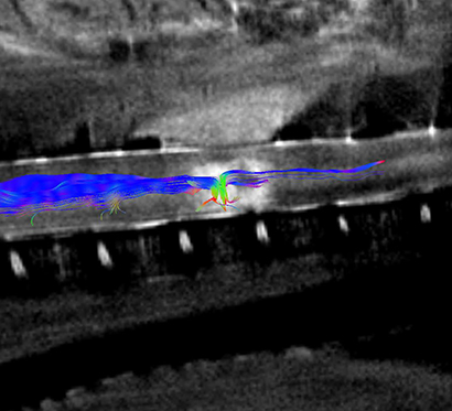
and after seeding a specific tract to visualize the status of the injury (bottom). Treatment progress can now be monitored in vivo.
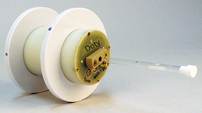
Both coils were tuned to 1H, for use with a 7 T small animal imaging system. Images were processed with DSI Studio software (Fang-Cheng (Frank) Yeh, Dept of Neurological Surgery, University of Pittsburgh).
From Laboratory of Dr. Prodip Bose; Malcom Randall VA Medical Center, Gainesville, FL.
